The Asymptomatic MVD Patient
Dr Nuala Summerfield and Dr Brigite Pedro
VVS Cardiology Specialists

Mitral valve disease (MVD) is the most common acquired cardiac disease in dogs. There have been some fairly recent innovations in staging and treatment of MVD. Read on to find out more about the latest thinking in this area.
Molly is an 8 year old neutered mixed-breed dog, weighing 8kg. Her owners have no concerns about her health. She has presented for an annual vaccination and health check. Your physical examination reveals no abnormalities until you auscultate her chest, at which point you detect a 3/6 left apical systolic murmur. No such murmur was detect at last year’s booster.
Her owners report that she has good exercise tolerance (she runs with her owner three or four miles a day!), has no cough or any episodes of fast or laboured breathing.

What is the most likely diagnosis?
A loud left apical systolic murmur in a small breed dog is most likely due to mitral valve disease.
Mitral Valve Disease
Mitral Valve Disease is due to chronic myxomatous degeneration of the valve leaflets, resulting in progressive valvular incompetence. As the amount of mitral regurgitation increases over time, this leads to progressive enlargement (remodelling) of the left atrium and left ventricle due to chronic volume overload.
There are therefore different stages of the disease. Currently, the preferred staging system is that issued by the American College of Veterinary Internal Medicine (ACVIM):
- Stage A – A patient without any evidence of cardiac disease (no murmur, asymptomatic, no cardiac remodelling), but who is considered at a genetically high risk. This would, for example, include high risk breeds like Cavalier King Charles Spaniels.
- Stage B – Patients in which there is evidence of valvular cardiac disease (i.e. a heart murmur), but no clinical signs.
- It can be subdivided into:
- B1 – Audible murmur with regurgitation detectable on echocardiogram, but the heart is otherwise structurally normal.
- B2 – Audible murmur with regurgitation present, and there is evidence of cardiac remodelling leading to left heart enlargement evident on radiography or echocardiography.
- It can be subdivided into:
- Stage C – Valvular disease is present, and clinical signs of congestive heart failure (e.g. tachypnoea, dyspnoea, cough, exercise intolerance, ascites, etc) have developed.
- Stage D – Valvular disease and congestive heart failure are present, but the clinical signs are becoming refractory to standard treatment (loop diuretics, ACE inhibitors, spironolactone and pimobendan).
What’s your next step with Molly?
Ideally we want to confirm our diagnosis and identify the stage of the disease.
As a mixed breed dog, Molly would not be considered Stage A unless there was a particularly strong history of cardiac disease in first degree relatives – sire, dam, or siblings. As she is not exhibiting any clinical signs as yet, she would not be considered Stage C, let alone Stage D. Molly fits into one of the Stage B categories. However, we cannot know which of the B subcategories without further investigations.
Staging MVD is extremely important. The reason for this is the EPIC study. This showed that starting pimobendan in stage B2 delays the onset of congestive heart failure by, on average, 15 months and reduces the risk of cardiac-related death by a third.
Introducing pimobendan at Stage B1 is unlikely to be harmful, but the drug is expensive, and there is no known benefit from doing so. It is therefore very important to be able to distinguish between dogs in stages B1 and B2!
Diagnostic Tests
Ideally an echocardiography (heart ultrasound) should be recommended. This is a more sensitive test which will confirm your diagnosis and help with staging.
Thoracic radiographs can also be considered to investigate possible cardiomegaly, which will be helpful for staging the disease.
Radiography
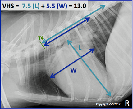
Evidence of cardiomegaly can usually be obtained easily through radiographs. Given the variable size and shape of the thoracic cavity, the use of a Vertebral Heart Score (VHS) is strongly recommended as a more sensitive and accurate measure of cardiac enlargement than simple observation or a subjective impression of “globularity”.

As you can see from the radiograph, Molly’s VHS is 13.0, which suggests marked cardiomegaly. Remember, the VHS is the length down the long axis of the heart plus the width across the short axis, measured in vertebrae. The upper limit for most dogs is 10.5, but for most purebreds, you may need to take into account breed features.
Echocardiography
Imaging of the mitral valve with a doppler-capable probe will give a clear indication of presence or absence of regurgitation. However, even without specialist training, it is quite easy to assess cardiac enlargement using most general practice ultrasound machines, using the LA:Ao ratio.
In transverse section, the diameter of the aortic root (the “Mercedes sign”) is measured, and compared to that of the Left Atrium (below and to the left on a standard projection). If the LA:Ao ratio is less than 1.6, the heart is considered normally sized; greater than 1.6 suggests cardiomegaly.


These echo images are from a dog with normal left atrial and left ventricular chamber size, consistent with Stage B1 MVD.
LA= Left Atrium, LV= Left Ventricle, Ao= Aorta


These echo images are from Molly and show significant left atrial and left ventricular chamber enlargement, consistent with Stage B2 MVD
The left ventricular internal dimensions in diastole on M-mode (LVIDd) adjusted to body weight can also be used to assess if there is any remodeling. There are formulas and tables where you can check if your measurements are normal for a specific body weight.

So, what stage is Molly in, and do we need to do anything?
We have at least two separate indices (VHS and LA:Ao) that suggest that Molly’s heart has started to remodel in response to volume overload caused by the mitral regurgitation. She would therefore be classified as Stage B2, and would thus be a good candidate for starting pimobendan.
Ideally a re-check should be recommended within 6 months but close monitoring is required until then. With the progression of the disease, it is possible that Molly will end up developing congestive heart failure and if so, at that time, she will require optimisation of the ongoing treatment. The owners should be instructed to start monitoring the resting respiratory rate at home and to contact you if this starts increasing over time.
Take home message:
- Mitral valve disease is the most common cause of heart murmurs in adult-middle aged and older small breed dogs.
- It is important to understand if there is cardiac remodelling (chamber enlargement) as this will affect prognosis and treatment options.
- Echocardiography and/or thoracic radiographs are important tools in the staging of mitral valve disease.
- The EPIC study showed that using pimobendan in dogs with mitral valve disease in stage B2 will delay the development of CHF and reduce the risk of cardiac death.
- Showing owners how to count the sleeping RR at home is an important tool for monitoring patients with stage B2 mitral valve disease.
Cardiology cases can feel overwhelming to deal with in general practice, but VVS’ friendly world-class cardiologists are on hand to support you and to enable you to bring outstanding clinical care to your patients and reassurance to their owners. Bring the specialist to you and your patient with the help of the VVS service. Get in touch to speak to us further about trying this unique service in your practice!
