

Dr Daniel Calvo Carrasco
LV CertAVP (ZM) DipECZM (Avian) MRCVS RCVS
Recognised Advanced Practitioner in Zoological Medicine EBVS® European Veterinary Specialist in Avian Medicine and Surgery
Lily, a 2-year-old, neutered, female, fully vaccinated, lop-eared rabbit is presented for a recurrent episode of lethargy and anorexia.
On at least two previous occasions your patient has responded to symptomatic treatment with meloxicam, prokinetics and syringe feeding. The owners are keen to investigate the cause of these episodes.
Q: What do you do first?
A: Full clinical examination
- Examine all body systems, but pay particular attention to teeth, gastrointestinal tract, cardiovascular and respiratory systems and ears. It may be necessary to sedate or anesthetise your patient to complete a thorough clinical examination.
- Manage your client’s expectations. Clients often underestimate both the time, equipment and cost involved in working up such cases. Let them know the options, but also the time frame and likely costs and what you hope to achieve.
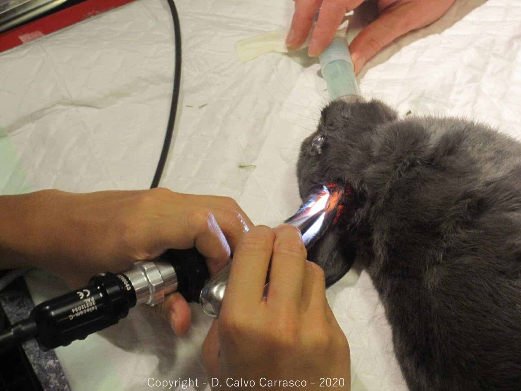
Q: Clinical examination is unremarkable, other than a small lump at the base of one ear. Otoscopic examination is difficult as the canal is quite stenotic, and the rabbit is resistant to otoscopy. What do you do next?
A: Lop-eared rabbits are prone to chronic external otitis. This preponderance for otitis is linked to the anatomy of our lop-eared patients and it is likely that this condition is under diagnosed.

In lop-eared breeds, there is gap of 3 to 5 mm between the annular cartilage and the auricular cartilage. It is this lack of continuous cartilage in the external ear canal that causes the ear to fold over (lop), creating a kink or a hinge in the ear canal. This hinge prevents the rabbit’s normal waxy ear secretions from draining adequately. As a result, these secretions accumulate and become an ideal media for naturally occurring bacteria and yeast to proliferate.
Affected rabbits may be asymptomatic, but as the condition progresses, they may start to display symptoms associated with discomfort (inappetence, lethargy, reduced faeces etc). Often affected rabbits will be presented in gut stasis. Gut stasis is a clinical presentation (rather than a disease in itself) and is caused by anything that causes the rabbit pain or stress. Some rabbits may go on to develop middle ear disease and may or may not present with a head tilt. Some rabbits may develop lytic lesions in the bulla and may develop facial paralysis.

Q: With all of this in mind, what would the next steps involve?
A: Next steps would involve the following:
- Sedating or anaesthetising your patient.
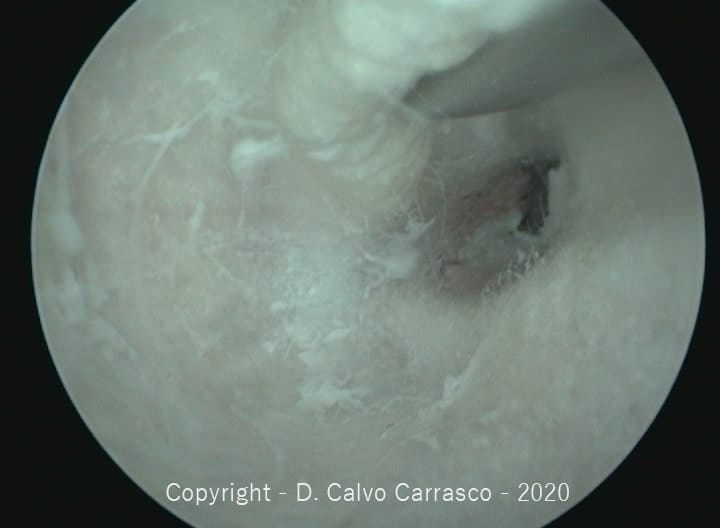
- To achieve a thorough otoscopic and/or endoscopic examination
- To take samples/swabs from the ear canal.
- To flush the ear canal. Note: if a diverticulum is present then problems tend to recur despite flushing.
- To aspirate the ear mass.
- To obtain a blood sample (it is not always necessary for rabbits to be anaesthetised for blood sampling, but it makes sense to do this if the patient is anaesthetised anyway, to minimise stress to them).
- Submit blood for Full blood count, haematology and biochemistry, plus E cuniculi serology.
- E. cuniculi serology is useful but interpreting the results can be challenging. IgG and IgM should be measured. 50% of the rabbit population is seropositive to IgG. Paired serology may be necessary to determine the relevance of positive results. Interpretation is complex and the diagnosis will depend on the serology data, combined with clinical findings. This all needs to be explained to owners who may be expecting a “quick result” from blood tests.
- Advanced Imaging. If you have access to advanced imaging modalities, then it may be possible to undertake this at the same time. However, bear in mind length of anaesthetic for these patients and consider undertaking imaging another day once you have the results of your blood work etc to assist with decision making.

Q: Bloods results indicate a mild anaemia, compatible with chronic disease. White blood cells are normal. Does this rule out an infectious process?
A: No. Rabbits with ear abscesses often have a normal WBC count, as the infections is localised to the chronic abscess.
Q: What imaging would you now do?
A: If you have the option of CT, then this is the optimal imaging modality for these cases. A CT scan allows you to explore the entire ear canal, including assessing any bony changes and involvement of concurrent dental and/or respiratory problems.


If you do not have access to a CT scanner, then radiography may be useful but can be difficult to interpret. Latero-20° ventral-laterodorsal oblique and rostro-30° ventral-caudodorsal projections are reported to be the most diagnostic for dogs and cats with otitis media. However, in rabbits the dorsoventral view is considered a better view in cases of suspected middle ear disease. The rostro-caudal projection can be useful too.

In Dr Calvo Carrasco’s experience it can be difficult to achieve a diagnosis with radiography. Unless one bulla is clearly affected and the other is not, then misdiagnosis is a real risk if relying on radiographic images alone.
Ultrasonography is of limited use due to the bony skull in this region
The rabbit in this case underwent a CT scan which confirmed a chronic external otitis with a diverticulum or pocket of pus (often incorrectly referred to as an ear base abscess)

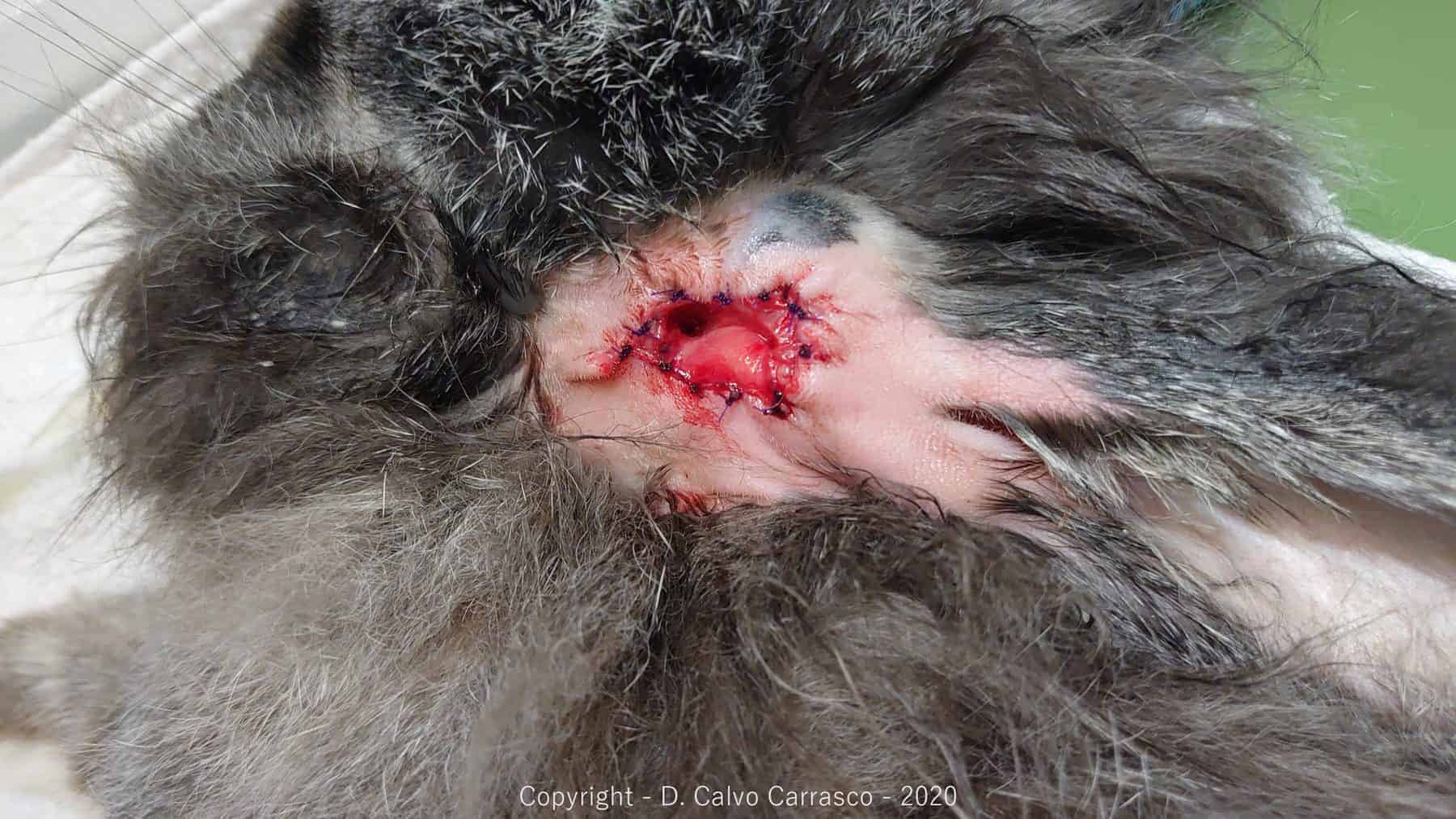
Q: What is the optimal treatment for this patient?
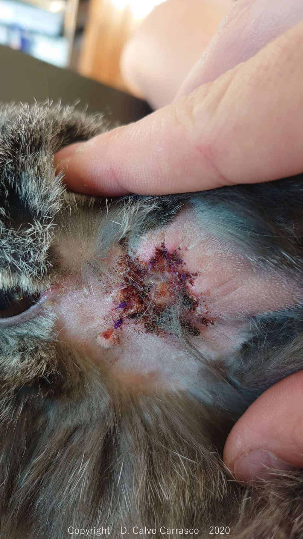
A: This patient underwent a partial lateral wall resection and made a full recovery. She is still doing well.
In Dr Calvo Carrasco’s experience, medical management is not usually successful when a diverticulum has formed. Although a diverticulum may remain unchanged for long periods of time, it can (and often does) progress to middle ear disease. In cases where there are ongoing clinical signs, surgical options tend to have a better long-term outcome.
Dr Calvo Carrasco recommends to fully excise abscesses in rabbits; or when this is not possible, to marsupialize them. This surgery allows excision of the affected area, while the permanent stoma prevents accumulation of secretions. Initial post-operative care is important to prevent the stoma from closing, but these patients then often require less ear cleaning than clinically well lop-eared rabbits.
Clinical decision making can be difficult in such patients, but the long-term benefits often outweigh the short-term challenges to the rabbit from this type of surgery. While surgery is invasive, this technique involves only skin, subcutaneous tissue and the epithelium of the ear canal, and if managed properly can improve patient welfare for the longer term.
If you would like assistance with your decision making, to ensure a holistic approach, combining excellent clinical care for your rabbit patients and ensuring optimal rabbit welfare, please get in touch with Dr Calvo Carrasco via the VVS website.

Q: What would the treatment options have been if this patient had been found to only have external otitis?
A: Medical management via otoscopic ear flush and topical treatment would be an acceptable first line treatment for a straight forward case of otitis externa, but owners should be made aware that this condition tends to recur as soon as they stop cleaning the ears. Therefore, partial lateral wall resection should also be considered as a treatment option even in milder, but recurrent cases. When the diverticulum has already appeared, medical management tends to be unsuccessful.
Q: What would be the treatment option if severe middle ear disease was diagnosed.
A: A partial ear canal ablation and a lateral bulla osteotomy (PECALBO)

Q: What could you do to minimise ear problems in lop-rabbits owned by your clients generally?
A: To prevent these rabbits becoming “ear patients”, it is a good idea to advise all owners of lop-eared rabbits to clean their ears regularly. It is important to demonstrate how to do this safely and effectively.